Not All MRIs Are Created Equal
July 09, 2024
Content created for the Bezzy community and sponsored by our partners. Learn More

Photography by Cavan Images/Getty Images
Magnetic resonance imaging (MRI) remains the gold standard for diagnosing MS and monitoring disease activity. But MRIs can differ in the type of experience and image clarity they offer.
The MRI. It is the most sensitive, noninvasive device currently available to image the brain and spinal cord.
For those of us living with multiple sclerosis (MS) and anyone undergoing tests for a possible diagnosis, the MRI allows our doctors a glimpse into the central nervous system — a chance to identify and monitor disease activity.
It is the most effective tool in the MS specialist’s shed. The one that neurologists turn to every day.
Indeed, some 90% of patients have their MS diagnosis confirmed by an MRI. And according to 2021 guidelines for people with MS, patients should have an MRI scan 3 to 6 months after starting new medications and then every year (or slightly less frequently following a few years of treatment if disease activity is stable).
All said, when you have MS, having MRIs becomes part of your life.
But what you may not know is that not all MRIs are the same.


MRI basics
A few quick facts about MRIs before we go deeper:
- Magnetic resonance imaging (MRI) uses magnetic fields and radio waves to measure water content in body tissue. As myelin — the fatty coating that protects nerves in our brain and spine — repels water, the MRI detects MS lesions where myelin tissue is holding more water (and is therefore damaged).
- Some MRIs use gadolinium. This is a contrast agent that can be injected into a blood vessel to help determine whether active inflammation is occurring.
- There are two main types of MRI:
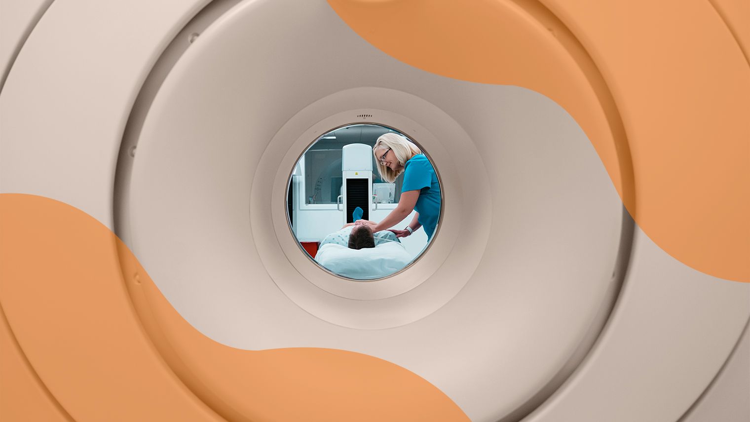
- Closed MRI resembles a big donut. The person lies on a table and is rolled into the “donut” for the MRI scan.
- Open MRI looks more like a sandwich. The magnets are positioned above and below the scanning table.
Not an open and closed case
Those of us who have been inside the bowels of a closed MRI can readily recall the experience. With thunderous clangs, clicks, whirrs, and beeps, it is loud (even with earplugs)!
And with our arms pinned to our waists, our hips hugging our legs together, and our heads locked into a helmet of sorts — called an MRI brain coil — it is tight!
While not enjoyable, for most people, the closed MRI experience is not terribly bothersome. But that’s just the case for some of us.
For people with obesity or claustrophobia, closed MRIs can feel like torture.
Small spaces, big issues
The dense constraints of the closed MRI pose very real complications for the more than 4 in 10 Americans who have obesity.
People with obesity face greater risks of thermal burns from unintended contact with the bore (the MRI opening) and can expect additional setup processes and acquisition times than people without obesity.
For those with claustrophobia — the fear of being in confined spaces — a closed MRI can be a non-starter. With a narrow tunnel and a scanner ceiling just inches from the person’s head and face, there may be nothing more terrifying than a closed MRI.
Open MRIs open the options
For people with obesity and claustrophobia, a newer option — the open MRI — may be a welcome alternative.
And yet, the open MRI also has some limitations. Chief among these is that the images that open MRIs provide are not as clear as those taken by closed MRIs.
That’s because open MRIs use weaker magnets, and the magnets are positioned farther away from the person. In general, these two factors determine the clarity and degree of detail in the pictures.
If having more detailed images is essential, those with claustrophobia may be able to take a sedative drug before the closed MRI scan to help them remain calm. Mindfulness techniques may also help people tolerate a closed MRI.
More about magnets
While closed MRIs do provide clearer images than open MRIs, there isn’t a one-strength-fits-all protocol for closed MRIs either.
Magnet strength is measured in a unit called Tesla (T). Some closed models use a 1.5-T magnet, while others employ 3.0-T magnets. If you live in a region with multiple MRI facilities, you may want to call around to determine which ones use a 3-T strength machine.
To add more complexity to the mix, in 2017, the Food and Drug Administration (FDA) approved the use of a 7-Tesla MRI — a new, even more powerful closed scanner.
While the 7-T machine does seem to offer superior detection and staging of MS, it is predominantly used as a research tool and not for patient care. Still, scientists believe the use of these more effective machines is likely to expand as research continues to uncover their benefits.
One certainty
With all this talk about the differences between MRIs, one thing is definite: They remain our best way to diagnose and monitor MS.
Regardless of which scanner you prefer, it’s important to follow your neurologist’s guidance and get new imaging done regularly.
We’re lucky to live in a time when medicine — and technology — are bettering our ability to live our lives with MS… even if it also means living with the clangs, clicks, whirrs, and beeps of an MRI.
Medically reviewed on July 09, 2024
9 Sources


Like the story? React, bookmark, or share below:
Have thoughts or suggestions about this article? Email us at article-feedback@bezzy.com.
About the author